Home

vaňa vyvariť delegácia gradient echo hyperintense lem sklo Premýšľavý

GBM, pre-contrast MRI | Lurie Family Imaging Center

Neuro-ophthalmology Question of the Week: Magnetic Resonance Imaging —  Neuro-Ophthalmology
Neuro-ophthalmology Question of the Week: Magnetic Resonance Imaging — Neuro-Ophthalmology

View Image
View Image

Hyperintense Thrombus on

MRI Features of Intracerebral Hemorrhage Within 2 Hours From Symptom Onset  | Stroke
MRI Features of Intracerebral Hemorrhage Within 2 Hours From Symptom Onset | Stroke

Gradient echo sequence demonstrating the small punctate hemorrhage as... |  Download Scientific Diagram
Gradient echo sequence demonstrating the small punctate hemorrhage as... | Download Scientific Diagram

Hypointensities in the Brain on T2*-Weighted Gradient-Echo Magnetic  Resonance Imaging - ScienceDirect
Hypointensities in the Brain on T2*-Weighted Gradient-Echo Magnetic Resonance Imaging - ScienceDirect

Gradient echo (GE) axial T2 weighted MRI at the level of C5 showing the...  | Download Scientific Diagram
Gradient echo (GE) axial T2 weighted MRI at the level of C5 showing the... | Download Scientific Diagram

Intracellular vs extracellular methemoglobin mri - Questions and Answers  ​in MRI
Intracellular vs extracellular methemoglobin mri - Questions and Answers ​in MRI

Flow on GRE sequences - Questions and Answers ​in MRI

EPOS™
EPOS™

Significance of Susceptibility Vessel Sign on T2*-Weighted Gradient Echo  Imaging for Identification of Stroke Subtypes | Stroke
Significance of Susceptibility Vessel Sign on T2*-Weighted Gradient Echo Imaging for Identification of Stroke Subtypes | Stroke
T2*-weighted imaging - Wikipedia

EPOS™
EPOS™

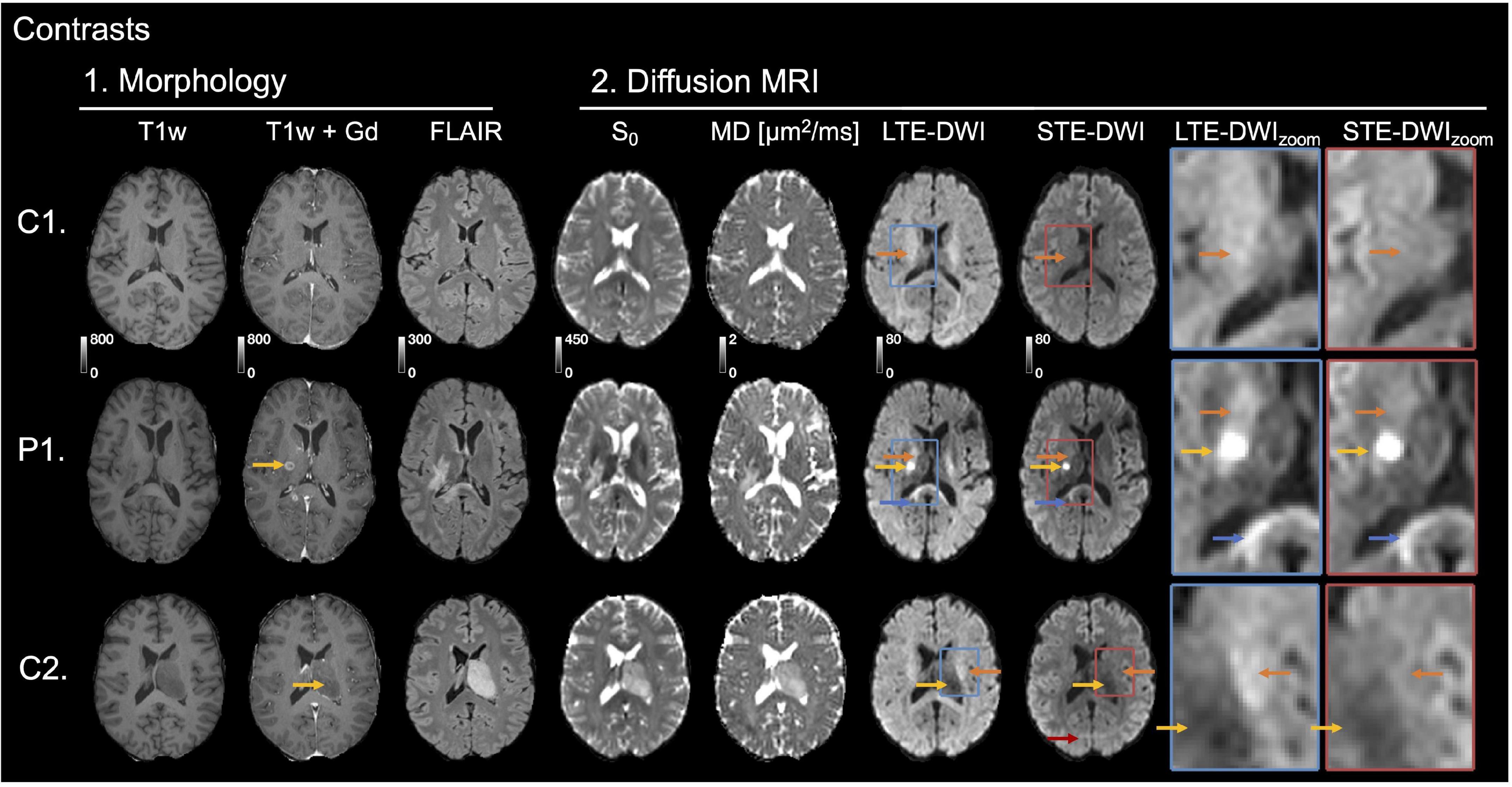
Frontiers | Separating Glioma Hyperintensities From White Matter by  Diffusion-Weighted Imaging With Spherical Tensor Encoding
Frontiers | Separating Glioma Hyperintensities From White Matter by Diffusion-Weighted Imaging With Spherical Tensor Encoding
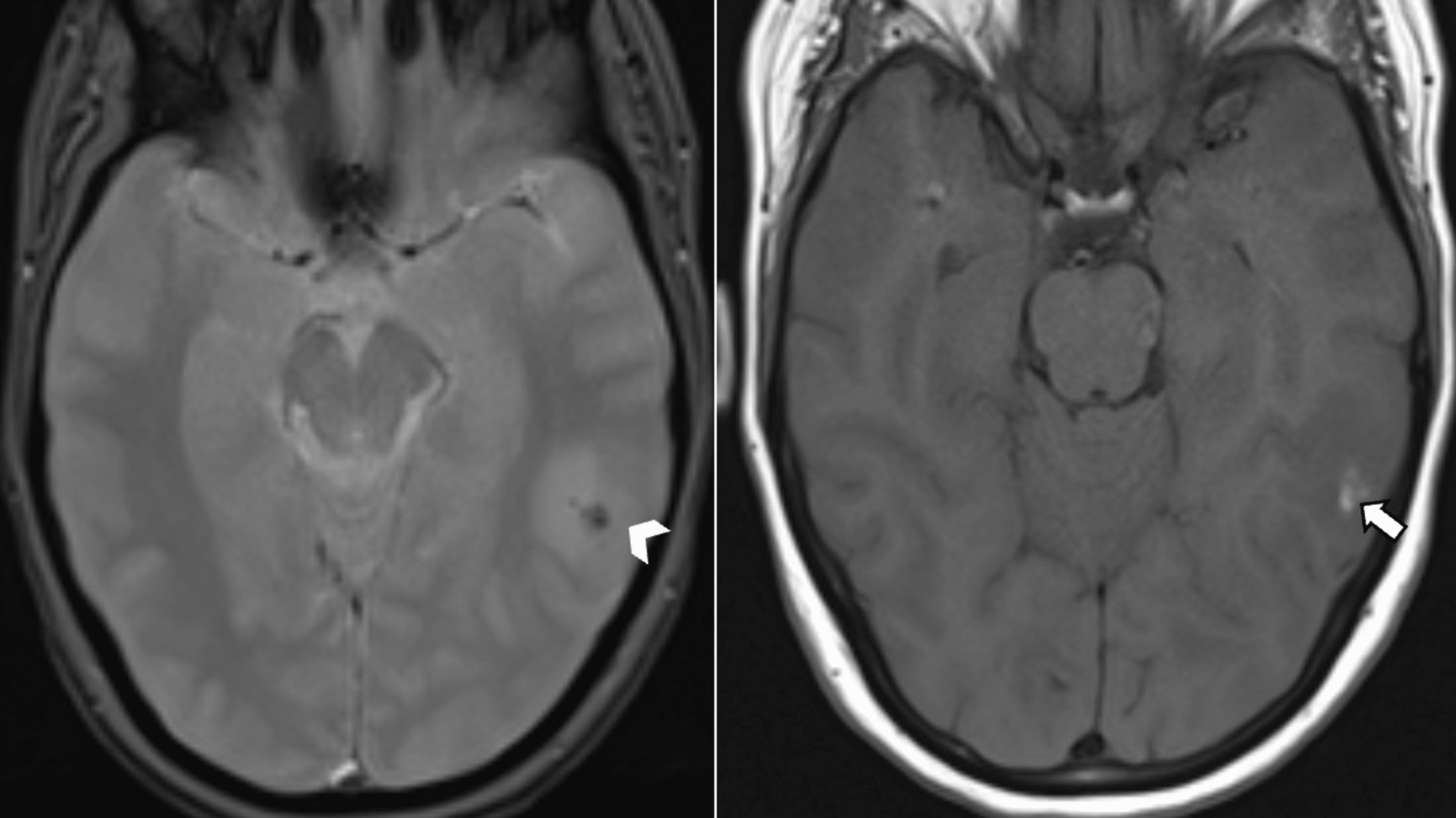
Cureus | Cerebral Vasculitis Revealing Systemic Sarcoidosis: A Case Report  and Review of the Literature | Article
Cureus | Cerebral Vasculitis Revealing Systemic Sarcoidosis: A Case Report and Review of the Literature | Article

fig 2. | Detection of Intracranial Hemorrhage: Comparison between Gradient- echo Images and b0 Images Obtained from Diffusion-weighted Echo-planar  Sequences | American Journal of Neuroradiology
fig 2. | Detection of Intracranial Hemorrhage: Comparison between Gradient- echo Images and b0 Images Obtained from Diffusion-weighted Echo-planar Sequences | American Journal of Neuroradiology

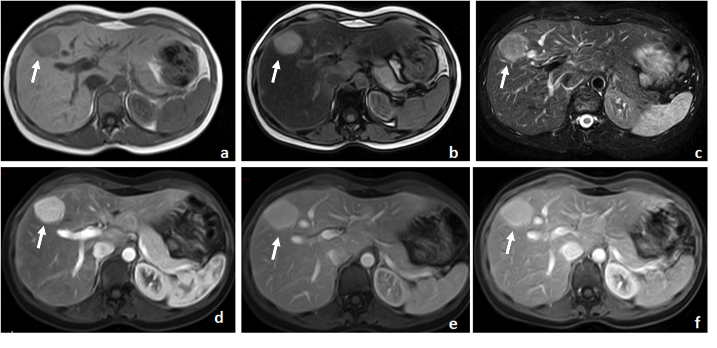
Understanding Body MRI Sequences and Their Ability to Characterize Tissues

Out of phase gradient echo T1 weighted sequence demonstrates... | Download  Scientific Diagram
Out of phase gradient echo T1 weighted sequence demonstrates... | Download Scientific Diagram

GRE T2-Weighted MRI: Principles and Clinical Applications

A) T1* gradient echo MRI. Images show abnormal low signal bilateral... |  Download Scientific Diagram
A) T1* gradient echo MRI. Images show abnormal low signal bilateral... | Download Scientific Diagram

Neurovascular Injury | Radiology Key

Automated detection of cerebral microbleeds on T2*-weighted MRI |  Scientific Reports
Automated detection of cerebral microbleeds on T2*-weighted MRI | Scientific Reports

Gradient echo images are superior to FLAIR in the detection of... |  Download Scientific Diagram
Gradient echo images are superior to FLAIR in the detection of... | Download Scientific Diagram

A gradient-echo T2*-weighted magnetic resonance image showing... | Download  Scientific Diagram
A gradient-echo T2*-weighted magnetic resonance image showing... | Download Scientific Diagram